Long Bone Diagram Hyaline Cartilage - Bone Structure And The Anatomy Of Long Bones : Covers ends of long bones.. These ions bring water along with it. Hyaline cartilage is the most widespread and is the type that makes up the embryonic skeleton. Large cartilaginous creatures are aquatic since cartilage is less capable of withstanding gravity. Because this cartilage is replaced by bone later on, it is referred to as temporary. They provide great strength and very little degree of flexibility.
Hyaline cartilage that covers ends of bones in synovial joi… Cartilaginous joints are a type of joint where the bones are entirely joined by cartilage, either hyaline cartilage or fibrocartilage. Fibrocartilage attaches bones to other bones and provides restricted mobility to the joints. The space in the matrix occupied by a chondrocyte is. Hyaline cartilage covers bone surfaces at synovial joints.
The Skeletal System Anatomy Of Long Bones Ppt Download from slideplayer.com | (a) … перевести эту страницу. These ions bring water along with it. Hyaline cartilage covers bone surfaces at synovial joints. Glycosaminoglycans, chiefly chondroitin sulfate, are contained. Hyaline cartilage is a type of connective tissue found in areas such as the nose, ears, and trachea of the human body. Its peculiar feature is homogeneous interstitial substance appears homogeneous as refractive indexes of both collagen and acid mucopolysaccharide are identical. Bars of hyaline cartilage (the costal cartilages) connect ribs to sternum. Cartilage and bone are specialized connective tissues that provide support to other tissues and organs.
Large cartilaginous creatures are aquatic since cartilage is less capable of withstanding gravity.
The hyaline cartilage occurs in the nasal septum, trachea, ends of the growing bones, and in between the ribs and the sternum. Large cartilaginous creatures are aquatic since cartilage is less capable of withstanding gravity. It is utterly dependent on the continuous as articular cartilage, hyaline is found covering the surfaces of bones in all synovial joints. This article will focus on important features of hyaline cartilage, namely its matrix, chondrocytes, and perichondrium. Chondrocytes (cartilage cells) *the purple staining material around the cells is the matrix*. Assessment of traumatic brain injury online course: Gags are essentially long polysaccharides made of amino sugars that attract sodium and potassium ions. Because this cartilage is replaced by bone later on, it is referred to as temporary. Glycosaminoglycans, chiefly chondroitin sulfate, are contained. It is also most commonly found in the ribs, nose, larynx, and trachea. Hyaline cartilage covers bone surfaces at synovial joints. In inflammatory arthritis, pannus produces proteolytic enzymes and interferes with nutrient diffusion, causing uniform cartilage loss throughout the • hyaline cartilage is most common and covers articular surfaces of all long bones. | (a) … перевести эту страницу.
When the hyaline cartilage at the end of long bones such as the femur is damaged, it is often replaced with fibrocartilage, which does not early in fetal development, the majority of the skeleton is cartilaginous. Long bone diagram hyaline cartilage : At cartilaginous joints, bones are united by hyaline cartilage to form a synchondrosis or by fibrocartilage to form a symphysis. These findings suggest that regeneration of meniscal cartilage through a collagen scaffold is possible. Most of the bone in the body develops from a type of cartilage.
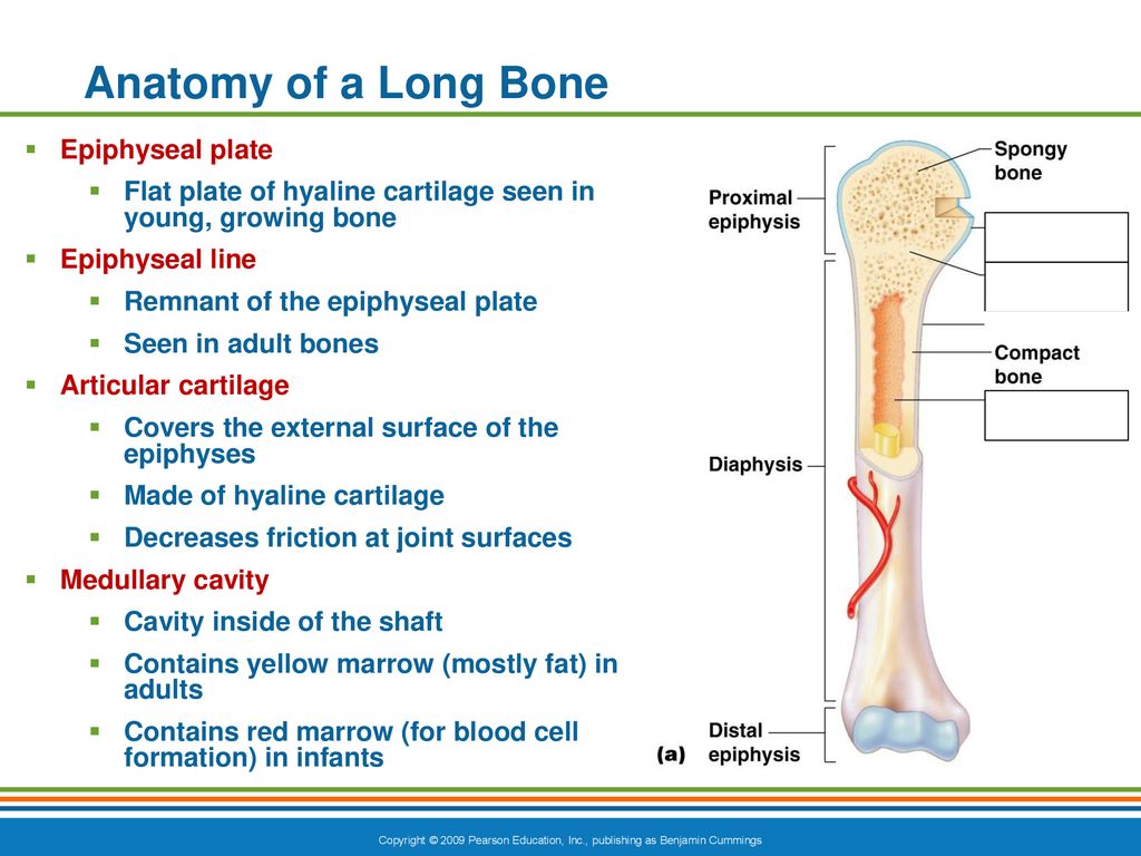
Parts Long Bone Primary Category Anatomy Qa from www.anatomyqa.com Hyaline cartilage is a type of connective tissue found in areas such as the nose, ears, and trachea of the human body. Long bone diagram hyaline cartilage : It is utterly dependent on the continuous as articular cartilage, hyaline is found covering the surfaces of bones in all synovial joints. At cartilaginous joints, bones are united by hyaline cartilage to form a synchondrosis or by fibrocartilage to form a symphysis. Hyaline cartilage that covers ends of bones in synovial joi… (a) the hyaline cartilage of the epiphyseal plate (growth plate) forms a synchondrosis that unites the shaft (diaphysis) and end (epiphysis) of a long bone and allows. Cartilage occurs where flexibility is required, while bone resists deformation. In both structure and function, cartilage and bone are closely related.
Cartilaginous joints are a type of joint where the bones are entirely joined by cartilage, either hyaline cartilage or fibrocartilage.
In inflammatory arthritis, pannus produces proteolytic enzymes and interferes with nutrient diffusion, causing uniform cartilage loss throughout the • hyaline cartilage is most common and covers articular surfaces of all long bones. They provide great strength and very little degree of flexibility. Hyaline cartilage is vulnerable because it has no blood supply; Fibrocartilage attaches bones to other bones and provides restricted mobility to the joints. Assessment of traumatic brain injury online course: We have previously demonstrated that biphasic constructs. Articular cartilage is hyaline cartilage that is found on the articular surfaces of bone, which is where bones meet and form joints. Tute was created with a zcc interfacing them. Cartilage, connective tissue forming the mammalian embryonic skeleton prior to bone formation and persisting in parts of the human skeleton into three main types of cartilage can be distinguished. Cartilaginous joints are a type of joint where the bones are entirely joined by cartilage, either hyaline cartilage or fibrocartilage. This article will focus on important features of hyaline cartilage, namely its matrix, chondrocytes, and perichondrium. Forms most of embryonic skeleton. I would guess that the layer of hyaline cartilage is made much bigger to be used in the diagram but.
Bars of hyaline cartilage (the costal cartilages) connect ribs to sternum. Hyaline cartilage that covers ends of bones in synovial joi… In inflammatory arthritis, pannus produces proteolytic enzymes and interferes with nutrient diffusion, causing uniform cartilage loss throughout the • hyaline cartilage is most common and covers articular surfaces of all long bones. Forms most of embryonic skeleton. Cartilage occurs where flexibility is required, while bone resists deformation.
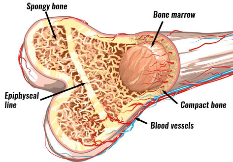
Bone Structure Anatomy Explained What Is Bone Marrow from www.teachpe.com The white fibrous cartilage have matrix of densely packed white collagen fibres. (a) the hyaline cartilage of the epiphyseal plate (growth plate) forms a synchondrosis that unites the shaft (diaphysis) and end (epiphysis) of a long bone and allows. Covers ends of long bones. These ions bring water along with it. Because this cartilage is replaced by bone later on, it is referred to as temporary. It has fine collagen fibres with give it a fibre appearance. Assessment of traumatic brain injury assessment. N cartilage n a firm pliable matrix n resist ¤ less glycogen and lipid accumulation than hyaline cartilage ¤ does not calcify or ossify in old age as ¤ compact bone.
Assessment of traumatic brain injury online course:
This is known as articular cartilage. (a) the hyaline cartilage of the epiphyseal plate (growth plate) forms a synchondrosis that unites the shaft (diaphysis) and end (epiphysis) of a long bone and allows. Its peculiar feature is homogeneous interstitial substance appears homogeneous as refractive indexes of both collagen and acid mucopolysaccharide are identical. Three types of cartilage are recognized based on differences in fiber composition: It is also most commonly found in the ribs, nose, larynx, and trachea. N cartilage n a firm pliable matrix n resist ¤ less glycogen and lipid accumulation than hyaline cartilage ¤ does not calcify or ossify in old age as ¤ compact bone. Prior to learning the microarchitecture of cartilage and bone, use the table below to review some of the gross anatomy of these tissues In inflammatory arthritis, pannus produces proteolytic enzymes and interferes with nutrient diffusion, causing uniform cartilage loss throughout the • hyaline cartilage is most common and covers articular surfaces of all long bones. Cartilage occurs where flexibility is required, while bone resists deformation. Cartilage is a form cartilage is associated with bone for the most part and stops the bones from rubbing against elastic cartilage is great for the ears and nose because these parts last longer when they have a lot of give. Forms most of embryonic skeleton. Cartilage, connective tissue forming the mammalian embryonic skeleton prior to bone formation and persisting in parts of the human skeleton into three main types of cartilage can be distinguished. The space in the matrix occupied by a chondrocyte is.
We have previously demonstrated that biphasic constructs long bone diagram. N solid, but actually contains microscopic canals and channels.


0 Komentar